The Fish Atlas.

Explore seven Utah fish species inside and out: 3D exteriors, illustrated organs and skeletons, feeding sequences, diet and source-backed science.

New: four-step feeding animation. Follow food and water through the fish. Play, pause, or choose a step in the illustrated cutaway.

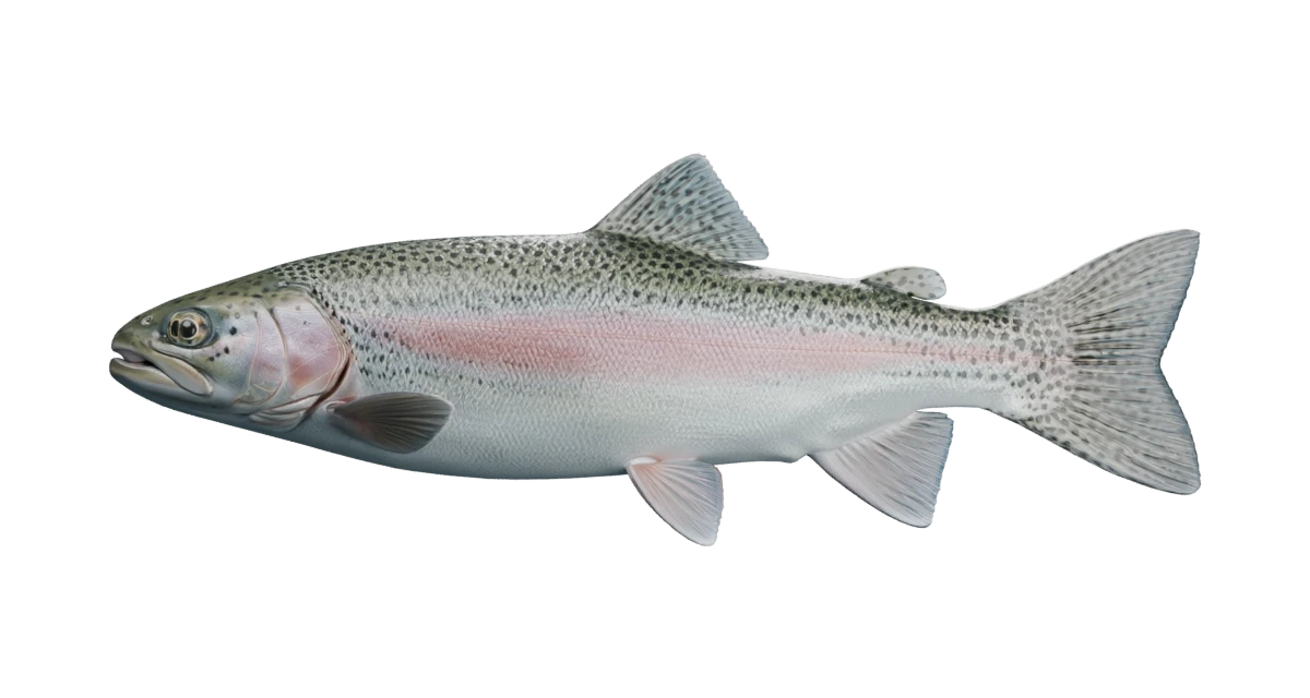
Illustrative rainbow trout exterior
Rainbow trout (Oncorhynchus mykiss). Illustrative exterior reconstruction with sourced organ and bone explanations.

Browse species

A closer look

Gill cover

Operculum

Shields the delicate gills. This bony cover participates in moving water through the gill chamber. Gas exchange happens at the gills underneath.

The curved plate behind the trout’s eye is the cover, not the gills themselves.

Sources: South Carolina DNR · Fish anatomy; Western Australia fisheries · Fish anatomy

Source-checked 2026-09-06. Independent anatomical review pending.

Lateral line

Detects nearby water movement. Small sensory organs along the flank respond to water motion. The highlighted line marks their general course, not a detection radius.

The lateral line and the rainbow’s pink stripe are different features, even where they lie close together.

On the water: Water movement is a sensory cue. It does not establish that a particular vibrating lure works better.

Sources: Western Australia fisheries · Fish anatomy; Utah DWR · Fish identification in the 2023 guidebook

Source-checked 2026-09-06. Independent anatomical review pending.

Tail fin

Caudal fin

Helps propel the fish. Sideways body bends and tail sweeps push against water to produce forward movement.

Small dark spots extend onto the rainbow trout’s tail.

Identification: Compare tail spotting with body pattern and other features; color alone can be misleading.

Sources: South Carolina DNR · Fish anatomy; Utah DWR · Fish identification in the 2023 guidebook; NOAA Fisheries · Fun facts about fascinating fish

Source-checked 2026-09-06. Independent anatomical review pending.

How this fish works

Built to move

The tail helps drive the fish forward. The other fins contribute to balance, braking and steering. Play the swimming illustration to follow their movement, or select each fin at your own pace.

Learn with the rainbow trout

How it breathes

Mouth and gill-cover movements move water across the gills. Dissolved oxygen crosses into the blood there. Routine ventilation can happen without eating. During suction feeding, a trout rapidly expands its mouth to draw prey inward.

Learn with the rainbow trout

More than meets the eye

Eyes gather visual information, nares sample odors, and the lateral line responds to water movement. These are different ways of sensing the surroundings. The highlighted line is a location, not a map of how far a fish can detect your lure.

Learn with the rainbow trout

Find this fish

A species listed in a water guide is not evidence of recent stocking. Compare the rainbow trout fishing guide, Utah water guides, and dated stocking records.

Sources & illustration notes

  1. South Carolina DNR · Fish anatomy

    External structure names, fins, smell and gill-cover function. General fish biology; not a rainbow trout dissection.

  2. Western Australia fisheries · Fish anatomy

    Paired fins, skin and scales, lateral line and gill physiology. Text used as reference; artwork is not reproduced.

  3. Utah DWR · Fish identification in the 2023 guidebook

    Rainbow trout exterior and identification. Identification reference only; use current DWR rules when planning a trip.

  4. U.S. Fish & Wildlife Service · Rainbow trout

    Scientific name and freshwater rainbow trout / steelhead life histories.

  5. Bureau of Land Management · Junior Explorer fish handbook

    Introductory explanation of water entering the mouth and passing out over gills.

  6. NOAA Fisheries · Fun facts about fascinating fish

    Body, tail and paired fins in fish locomotion.

  7. Koll et al. (2020) · Gene profiling in the adipose fin of salmonid fishes

    Rainbow trout evidence supporting a possible flow-sensing function; not proof of a complete behavioral mechanism.

  8. California Department of Fish and Wildlife: trout internal anatomy

    Trout organ locations and relationships; a reference for the simplified cutaway, not a license for the artwork.

  9. Oregon State University / ODFW: Salmonid Dissection Handbook

    Figure 2: salmonid organ arrangement and digestive tract; gill rakers and organ identification. Used for anatomical layout, not all physiological statements in the handbook.

  10. Huchzermeyer: Introduction to anatomy and physiology of fish

    Trout organs, gill countercurrent exchange, circulation and freshwater salt/water balance. Veterinary training material hosted by WOAH.

  11. Provini & Van Wassenbergh (2018): suction-feeding hydrodynamics

    General mechanics of mouth expansion and water outflow. The study models sunfish, not rainbow trout; it does not validate the timing of this illustration.

  12. Jimenez & Camp (2023): vertebral curvature during trout feeding

    Measured body-muscle deformation and vertebral bending during rainbow trout feeding strikes.

  13. Sugiura (2026): skeletal tissues in rainbow trout

    Normal control specimens illustrate vertebrae, abdominal ribs, neural/hemal arches and caudal supports. The exhibit does not depict the phosphorus-deficient specimens.

  14. Verdile et al. (2020): the rainbow trout intestine

    Rainbow trout digestive tract, pyloric caeca and regional digestive/absorptive function.

  15. Maine Inland Fisheries & Wildlife: rainbow trout

    Diet changes with fish size, habitat and season. Maine fishing regulations and regional size patterns are not applied to Utah.

  16. USGS: Oncorhynchus mykiss species profile

    Rainbow trout feeding ecology and foods including invertebrates, fish eggs and small fish.

Reference fish and river artwork generated with Higgsfield; exterior mesh generated with Meshy 7 through fal. Optimized, annotated and animated in Blender for Utah Stocked Fish, September 2026. Internal and skeletal illustrations generated with Higgsfield GPT Image 2.5 for this exhibit, September 2026. Generated using the connected provider accounts. No third-party Creative Commons license or expert endorsement is asserted.

The 3D exterior and the fixed-side internal illustrations are simplified artistic reconstructions, not scans. Internal views show selected organ regions; overlapping tissues are separated for clarity. Bone counts, small skeletal elements, vessels, gonads and microscopic gill detail are not represented precisely. Flow arrows explain direction, not measured flow or feeding speed. Biological explanations are source-checked; independent expert review of the illustrations and motion is pending.