Crayfish on the bottom
Crayfish are important prey in many smallmouth waters. Rocky habitat offers both hiding places for prey and feeding opportunities for bass.
Sources: Minnesota DNR — Smallmouth bass biology and habitat
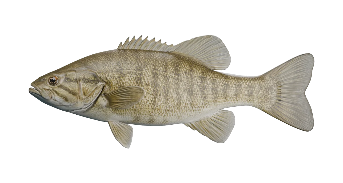
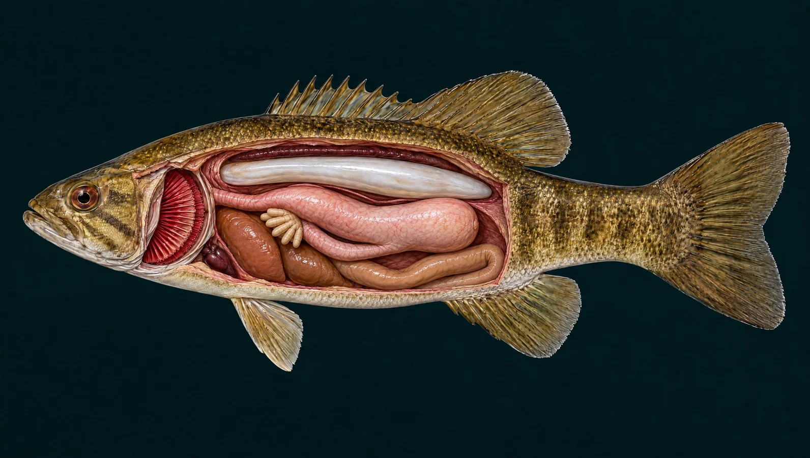
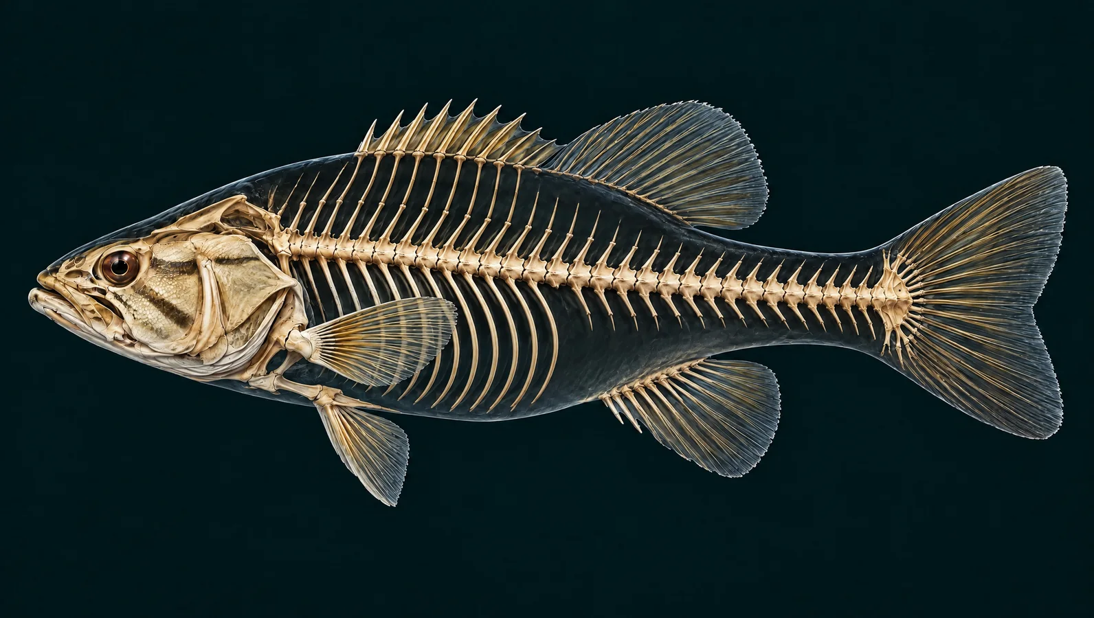
Bronze sides, a compact mouth, and fins ready for quick changes of direction. Explore the smallmouth bass from its rocky feeding grounds to the bones, gills and digestive organs that keep it moving.
New: four-step feeding animation. Follow food and water through the fish. Play, pause, or choose a step in the illustrated cutaway.
Fixed-side artist illustrations, not scans. Organ regions and skeletal details are simplified; independent expert review is pending.
The stages separate events for teaching; their timing can overlap in a real fish.
Smallmouth combine movement toward prey with suction. Experiments found different combinations when taking bottom prey and prey swimming in open water.
Sources: Sejdic (2016) — Prey capture and suction in smallmouth bass
Water passes through the gill region and leaves behind the gill cover. Captured food stays in the mouth and throat for handling. The arrows show general direction, not every fluid motion of a strike.
Sources: Evans, Piermarini & Choe (2005) — The multifunctional fish gill; Sejdic (2016) — Prey capture and suction in smallmouth bass; Camp & Van Wassenbergh (2025) · A mechanical perspective on suction feeding in fishes
Food is swallowed through the esophagus. It does not go through the gill filaments: the respiratory route and digestive route separate behind the mouth.
Sources: Australian Museum — Parts of a fish; FAO — Digestion in teleost fishes; Camp & Van Wassenbergh (2025) · A mechanical perspective on suction feeding in fishes
Stomach, pyloric caeca and intestine process the meal. The anatomical layout is simplified and the animation compresses a slow process into a few seconds.
Sources: FAO — Digestion in teleost fishes; University of Texas at Austin — Fishes of Texas: Micropterus dolomieu
Crayfish are important prey in many smallmouth waters. Rocky habitat offers both hiding places for prey and feeding opportunities for bass.
Sources: Minnesota DNR — Smallmouth bass biology and habitat
Smallmouth also eat small fish. Their feeding movements can shift when pursuing a swimming target rather than taking food from the bottom.
Sources: South Carolina DNR — Smallmouth bass; Sejdic (2016) — Prey capture and suction in smallmouth bass
Young smallmouth take zooplankton and aquatic insects; larger fish add more crayfish and fish. Local availability still matters.
Sources: South Carolina DNR — Smallmouth bass; U.S. Fish & Wildlife Service — Smallmouth bass ecological summary
Rocky shoals, drop-offs, river eddies and pools can connect bass with prey. These are habitat clues, not evidence that a particular lure must work.
Sources: Minnesota DNR — Smallmouth bass biology and habitat
Back to species guide · Interactive controls become available when JavaScript loads. Read all explanations below.
Takes in prey and water. The mouth cavity expands rapidly during a strike, drawing in nearby water and food.
With the mouth closed, the upper jaw usually ends below the eye rather than behind it.
Identification: Compare jaw position on a real fish with its mouth closed, alongside the dorsal-fin notch and body markings.
Sources: South Carolina DNR — Smallmouth bass; Sejdic (2016) — Prey capture and suction in smallmouth bass
Source-checked 2026-09-19. Independent anatomical review pending.
Detects light and movement. Vision contributes to locating prey and navigating around obstacles; light and water clarity affect what is visible.
The reddish-brown eye in this reconstruction is one variable feature, not a stand-alone identification test.
Sources: South Carolina DNR — Fish anatomy; South Carolina DNR — Smallmouth bass
Source-checked 2026-09-19. Independent anatomical review pending.
Admit water to the smell organs. These openings sample dissolved chemicals. Breathing water enters through the mouth rather than through the nares.
Tiny openings ahead of the eye are enlarged by the marker for visibility.
Sources: South Carolina DNR — Fish anatomy
Source-checked 2026-09-19. Independent anatomical review pending.
Operculum
Protects the gills and helps ventilate them. Movement of the mouth and gill-cover cavities helps move water over the respiratory surfaces.
Look behind the cheek markings for the rear edge of this bony flap.
Sources: South Carolina DNR — Fish anatomy; Evans, Piermarini & Choe (2005) — The multifunctional fish gill
Source-checked 2026-09-19. Independent anatomical review pending.
Detects nearby water motion. Sensory cells in canals respond to local water movement. This is not a sonar beam or a promise that a particular lure will work.
The canal crosses the bronze flank; the vertical bars are pigment, not sensory organs.
Sources: South Carolina DNR — Fish anatomy; South Carolina DNR — Smallmouth bass
Source-checked 2026-09-19. Independent anatomical review pending.
Helps stabilize the body; stiff spines support the fin. The front portion is supported by rigid spines. It remains joined to the soft-rayed portion behind it.
A relatively shallow notch joins the spiny and soft portions.
Identification: Use the shallow dorsal notch together with the smaller jaw and vertical body markings.
Sources: South Carolina DNR — Smallmouth bass; South Carolina DNR — Fish anatomy
Source-checked 2026-09-19. Independent anatomical review pending.
Provides a flexible surface for swimming control. Segmented rays support the rear dorsal membrane, which works with the other fins during movement.
It is broadly joined to the spiny portion. There is no adipose fin.
Sources: South Carolina DNR — Smallmouth bass; USGS — BEST field procedures, Appendix C anatomy
Source-checked 2026-09-19. Independent anatomical review pending.
Helps steer, brake and hold position. The paired fins behind the head make smaller adjustments while body and tail provide thrust.
Rounded pectoral fins help the fish control its position around rocky cover.
Sources: South Carolina DNR — Fish anatomy; University of Texas at Austin — Fishes of Texas: Micropterus dolomieu
Source-checked 2026-09-19. Independent anatomical review pending.
Helps control balance and orientation. These paired ventral fins assist in positioning and slowing the fish.
Bass have thoracic pelvic fins: they sit near the front, below the pectoral region, rather than halfway along the belly like a trout.
Sources: University of Texas at Austin — Fishes of Texas: Micropterus dolomieu; South Carolina DNR — Fish anatomy
Source-checked 2026-09-19. Independent anatomical review pending.
Helps keep the fish stable in motion. This unpaired fin lies on the underside behind the vent, working with the dorsal fin.
Its stiff leading spines and flexible rays are shown as broad regions; the generated model is not a fin-ray counting guide.
Sources: USGS — BEST field procedures, Appendix C anatomy; South Carolina DNR — Smallmouth bass
Source-checked 2026-09-19. Independent anatomical review pending.
Caudal fin
Transfers body movement into thrust. Side-to-side bending of the body drives the caudal fin against the surrounding water.
The shallowly notched tail supports acceleration and maneuvering between pools and rocky feeding areas.
Sources: FAO — Biological aspects of fish; University of Texas at Austin — Fishes of Texas: Micropterus dolomieu
Source-checked 2026-09-19. Independent anatomical review pending.
Protect the outer body. Overlapping scales form a flexible covering; the outer skin and mucus also help protect the fish.
Smallmouth have ctenoid scales with fine comb-like edges; those microscopic edges are not resolved here.
Sources: University of Texas at Austin — Fishes of Texas: Micropterus dolomieu; South Carolina DNR — Fish anatomy
Source-checked 2026-09-19. Independent anatomical review pending.
Exchange gases between water and blood. Thin lamellae provide exchange surfaces. Water and blood pass in opposite directions at these surfaces, helping oxygen move into the blood.
The red region marks the gill chamber; individual arches and microscopic lamellae are simplified.
Sources: Evans, Piermarini & Choe (2005) — The multifunctional fish gill; Australian Museum — Parts of a fish
Source-checked 2026-09-19. Independent anatomical review pending.
Pumps blood toward the gills. Blood travels from the heart to the gills, then to body tissues before returning to the heart.
The heart occupies a small ventral region just behind the gill apparatus; separate chambers are not modeled.
Sources: FAO — Biological aspects of fish; Australian Museum — Parts of a fish
Source-checked 2026-09-19. Independent anatomical review pending.
Processes nutrients and contributes bile for digestion. Absorbed nutrients reach this metabolic organ, which helps manage fuel and materials used throughout the body.
The liver is shown partly separated from neighboring gut tissue so the digestive route can be followed.
Sources: FAO — Digestion in teleost fishes; FAO — Biological aspects of fish
Source-checked 2026-09-19. Independent anatomical review pending.
Passes swallowed food from throat to stomach. The food passage runs behind the gill region into the stomach; it is separate from the water exit behind the gill cover.
The illustration exposes a short part of the passage, not the entire folded throat apparatus.
Sources: Australian Museum — Parts of a fish; FAO — Digestion in teleost fishes; Camp & Van Wassenbergh (2025) · A mechanical perspective on suction feeding in fishes
Source-checked 2026-09-19. Independent anatomical review pending.
Holds food and begins chemical digestion. The muscular stomach mixes a meal with digestive secretions before material enters the intestine.
A stomach and intestinal tract process animal prey. The tissue shape shown here is a broad reconstruction, not a specimen measurement.
Sources: FAO — Digestion in teleost fishes; University of Texas at Austin — Fishes of Texas: Micropterus dolomieu
Source-checked 2026-09-19. Independent anatomical review pending.
Add digestive and absorptive surface near the gut entrance. These blind-ending pouches connect near the stomach-intestine junction. They are part of the digestive system, not eggs or worms.
Smallmouth possess unbranched pyloric caeca. This cluster shows their general region, not a count.
Sources: University of Texas at Austin — Fishes of Texas: Micropterus dolomieu; FAO — Digestion in teleost fishes
Source-checked 2026-09-19. Independent anatomical review pending.
Continues digestion and absorbs nutrients. Material moves through the gut; usable nutrients enter the body, and undigested remains eventually leave through the anus.
The loop is shortened and separated for clarity. Food does not travel through the gills or swim bladder.
Sources: FAO — Digestion in teleost fishes; University of Texas at Austin — Fishes of Texas: Micropterus dolomieu
Source-checked 2026-09-19. Independent anatomical review pending.
Helps control buoyancy. Gas inside the bladder reduces the effort needed to maintain depth. It is not a stomach and does not receive food.
The bladder lies above the digestive organs. Its broad location is shown; gas-control vessels are omitted.
Sources: Western Australia DPIRD — Fish anatomy; Australian Museum — Parts of a fish
Source-checked 2026-09-19. Independent anatomical review pending.
Helps regulate internal water and ion balance. Excretion and water balance involve both the kidneys and gills; a freshwater bass must regulate exchange with its surroundings.
The thin dorsal strip marks the trunk-kidney region beneath the backbone, above the swim bladder.
Sources: Evans, Piermarini & Choe (2005) — The multifunctional fish gill; FAO — Biological aspects of fish; Australian Museum — Parts of a fish
Source-checked 2026-09-19. Independent anatomical review pending.
Supports the head and its feeding apparatus. A fish skull contains multiple linked elements; feeding involves coordinated movement rather than a single rigid head.
The smaller gape changes the feeding apparatus compared with a largemouth; this broad skull outline is illustrative.
Sources: Sejdic (2016) — Prey capture and suction in smallmouth bass; USGS — BEST field procedures, Appendix C anatomy
Source-checked 2026-09-19. Independent anatomical review pending.
Open the feeding aperture and grasp prey. Linked jaw elements move as the mouth opens. Fine teeth and small joint surfaces are not individually modeled.
Smallmouth can protrude the upper jaw during prey capture, but do not have the long gape of a largemouth.
Sources: Sejdic (2016) — Prey capture and suction in smallmouth bass; South Carolina DNR — Smallmouth bass
Source-checked 2026-09-19. Independent anatomical review pending.
Vertebral column
Supports the body while allowing it to bend. A series of vertebrae supports muscles and helps transmit movement along the fish.
Only the main column and broad projections are illustrated. Do not use this asset to count vertebrae or diagnose injuries.
Sources: USGS — BEST field procedures, Appendix C anatomy; FAO — Biological aspects of fish
Source-checked 2026-09-19. Independent anatomical review pending.
Support the body wall around the abdominal cavity. Slender ribs extend from the anterior trunk. Toward the tail, the arrangement changes to a narrower framework.
The rib region ends around the abdomen; small intermuscular bones are not precisely resolved.
Sources: USGS — BEST field procedures, Appendix C anatomy; Australian Museum — Parts of a fish
Source-checked 2026-09-19. Independent anatomical review pending.
Support the flexible membranes of the fins. Soft rays are segmented supports that can bend with fin movement, unlike the rigid front dorsal spines.
This marker highlights caudal rays. Branching and counts throughout the reconstruction are approximate.
Sources: USGS — BEST field procedures, Appendix C anatomy; University of Texas at Austin — Fishes of Texas: Micropterus dolomieu
Source-checked 2026-09-19. Independent anatomical review pending.
Pterygiophores
Anchor and support the dorsal fin. Internal support elements lie between the back muscles and the fin, transferring forces to its spines and rays.
These are slender fin supports, not a second backbone. Their exact articulations are simplified.
Sources: USGS — BEST field procedures, Appendix C anatomy
Source-checked 2026-09-19. Independent anatomical review pending.
A smaller jaw, bronze sides with vertical bars and a shallow dorsal notch distinguish the smallmouth's profile. Color alone varies too much to settle every identification.
Tail-driven acceleration works with the paired fins to control position around rocks and changing currents. The swimming animation illustrates a bend traveling toward the tail.
Water entering the mouth moves over the gills and exits behind the cover. At the tiny exchange surfaces, blood and water flow in opposite directions; oxygen crosses into blood.
Smallmouth alter the balance between forward movement and suction when taking different prey. Follow the four stages to separate water flow, swallowing and digestion.
The lateral line responds to nearby water motion, the eyes detect visual information, and the nares sample dissolved chemicals. These systems contribute different information; an illustrated signal is not a lure-performance test.
Food passes through the esophagus into a bent stomach. Pyloric caeca and intestine continue processing and absorption. Above them, the swim bladder contributes buoyancy; it is separate from the food route.
One vertebral column supports the body. Abdominal ribs flank the organ cavity, while skull elements, fin supports and rays enable movement. This illustration shows broad regions; individual bone counts and tiny joints are approximate.
Explore the species guide and connected Utah waters. Known presence helps plan a visit, while recent stocking is a separate record that needs its own date and source.
A species listed in a water guide is not evidence of recent stocking. Compare the smallmouth bass fishing guide, Utah water guides, and dated stocking records.
Basic fin, gill-cover, scale, smell and lateral-line functions.
Largemouth internal plates for broad organ and skeletal relationships; the separate whole-skeleton plate is Nile perch. Used as a factual reference, not reproduced artwork.
Verified largemouth organ and skeletal plate, Appendix C, page 61, credited to Hunn (1988). Comparative bass layout, not a smallmouth specimen or exact reconstruction.
Single-loop circulation, transport of nutrients and broad muscle function in bony fishes.
General stomach, intestine, liver and digestive anatomy; not species-specific measurements.
Gill lamellae, opposite directions of water and blood flow, oxygen diffusion, and ion/water regulation. The inset is a general teleost explanation.
Broad bony-fish structures, buoyancy and body form. Marine water-balance examples are not applied to these freshwater bass.
Jaw position, connected dorsal sections, bronze/vertical markings, early diet and habitat.
Crayfish, small fish and insects; rocky shoals, eddies and pools. Minnesota regulations are not presented as Utah rules.
Official abstract of Sejdic’s 2016 master’s thesis: high-speed experiments on smallmouth suction and forward motion with benthic versus pelagic prey. Full thesis was not read in this audit.
Smallmouth external anatomy, ctenoid scales, jaw teeth, thoracic pelvic fins, and presence of unbranched pyloric caeca.
Diet changes with growth and ecological effects of introduced populations; not recent stocking records.
Review of capture and transport, including gill-arch and pharyngeal structures. Some transport details remain unresolved; this is general evidence, not measured motion of the illustrated bass.
Species reference and separate internal illustrations generated with Higgsfield GPT Image 2.5; exterior mesh generated with Meshy 7 through fal. Prepared for Utah Stocked Fish, September 2026. Generated through the connected provider accounts. No Creative Commons license, specimen scan or expert endorsement is asserted.
Exterior geometry and fixed-side cutaways are artistic reconstructions. Internal views separate overlapping regions for teaching. Small bones, exact counts, tissue sizes, microvessels, gonads and microscopic gill geometry are not represented precisely. Animation and flow paths are illustrative, not measured kinematics. Independent anatomical review is pending. Organ placement is a source-grounded bass layout adapted to the smallmouth silhouette, not a dissection or scan of this individual.