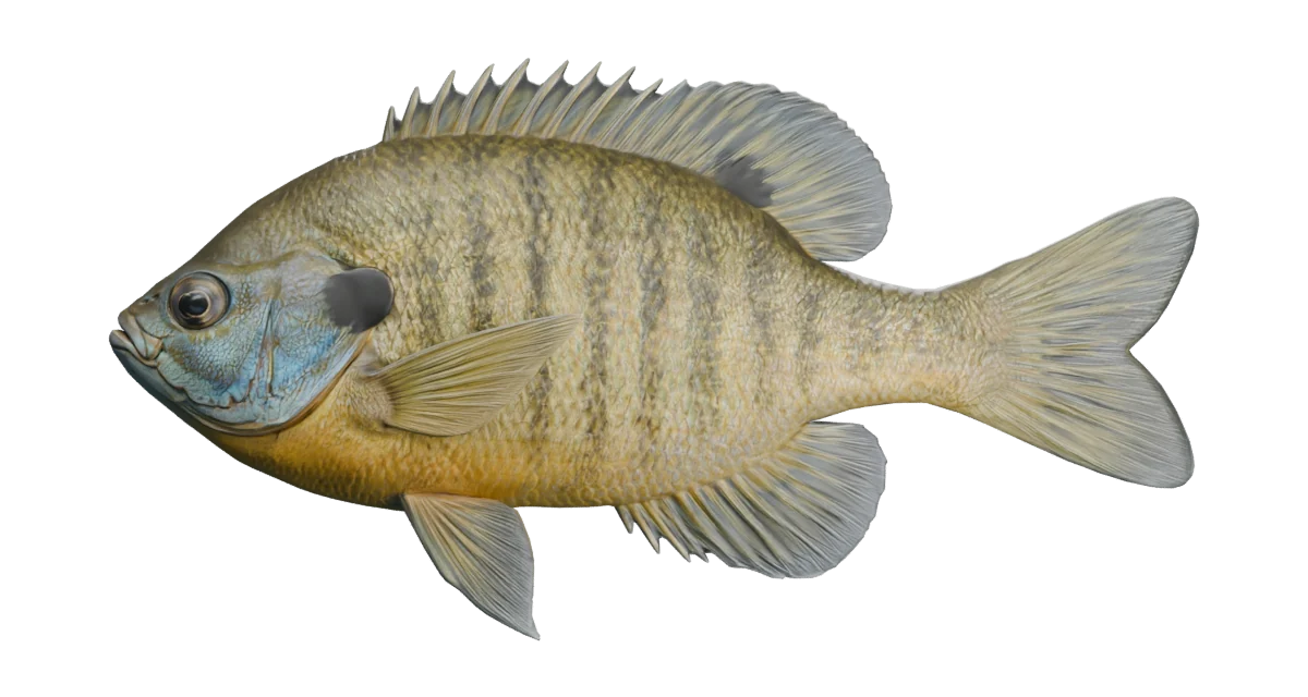
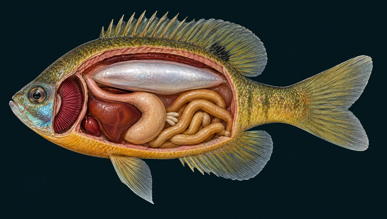
A small fish with remarkable control. Explore the bluegill's vivid blue cheek, tiny suction-feeding mouth and versatile fins, then look inside its deep body to follow breathing, digestion and buoyancy.
Bluegill (Lepomis macrochirus). Illustrative exterior reconstruction with sourced organ and bone explanations.
Inside the fish
Fixed-side artist illustrations, not scans. Organ regions and skeletal details are simplified; independent expert review is pending.
Selected organ regions, separated for clarity.
One gulp. Two routes.
The stages separate events for teaching; their timing can overlap in a real fish.
Expand the small mouth
A quick expansion of the mouth-and-throat space lowers pressure, drawing nearby water and small prey inward. The illustration slows a fast strike so you can inspect it.
Water passes the gill region and exits behind the covers. Gill rakers help retain food; movements of the mouth and throat then carry it toward the esophagus. The rakers themselves are not resolved in this cutaway.
The stomach begins digestion; the pyloric pouches and intestine contribute further digestive and absorptive surface. This stage takes much longer than the capture sequence.
Bluegill may also eat snails, small crayfish, fish eggs or very small fish. Mouth size and local food availability affect what an individual can capture.
Take dissolved oxygen from water. Water passes over thin exchange surfaces while blood flows through them. Oxygen enters the blood and carbon dioxide leaves.
The tall fan here shows the gill region. Microscopic lamellae are enlarged only in the separate teaching inset.
Carries food from the throat to the stomach. After prey is retained, it moves backward into the digestive tract while most water from capture leaves through the gills.
This short tube is distinct from the gill chamber even though both connect with the mouth region.
Supports senses and the feeding apparatus. Multiple head bones support movable jaws and the expanding mouth cavity.
Bluegill have many linked cranial bones. This guide covers their broad function; fine sutures and hidden throat bones are not individually illustrated.
Start with a deep, flattened body and a small mouth. Add the dark ear flap, blue cheek and connected spiny-and-soft dorsal fin. Together these features are more useful than any one patch of color.
Fine control between the stems
Bluegill coordinate several fins to maneuver. Pectoral fins can provide low-speed thrust and precise steering; the tail joins more strongly during active swimming and acceleration.
Water keeps oxygen arriving
Mouth and gill-cover movements keep water crossing the gills. The blood takes up dissolved oxygen there. In the enlarged teaching inset, follow opposing water and blood directions across a tiny exchange surface.
A tiny mouth makes suction
A rapid expansion draws nearby prey inward with water. Food is retained and transported into the digestive tract; water continues out through the gill openings. The numbered stages let you pause that sequence.
Spines and rays do different jobs
Compare the stiff supports at the front of the dorsal fin with flexible rays behind them. The backbone supports the deep body while allowing bending, and ribs frame the abdominal cavity.
Spot, approach, investigate
Vision helps a bluegill approach small moving prey. Smell and local water-motion sensing provide other information. The lateral line arches high on the flank; its highlight is a location, not a lure-detection range.
An efficient arrangement
The swim bladder lies above the digestive organs, with kidney tissue along the cavity roof. A curved stomach, short pyloric pouches and looping intestine process a meal below. Select each region to connect place and function.
Look closely at a familiar fish
Carry these features into the existing fishing guide and water pages. A familiar pond fish can be a careful observation lesson: compare the mouth, fins and flap before checking current access and dated stocking records.
Species identity, small mouth, black flap, joined dorsal regions, pointed pectoral fin and feeding ecology. Missouri regulations are not applied to Utah.
Bluegill organ identification, pyloric caeca, gills and fin locations. The handout's simplified liver/bile and heart-chamber statements are not adopted; physiology is checked separately.
Bluegill fin spines, segmented fin rays and micro-CT of pectoral supports; general bony-fish backbone framework. The overview skeleton is perch-like, not a species-specific whole bluegill scan.
Primary osteology of 100 specimens, including a whole-fish X-ray and bone/fin counts. Used to correct skeletal teaching and identify artwork errors. Source plate not reproduced; no public reuse license was established.
Species-specific reference and internal/skeletal illustrations generated with Higgsfield GPT Image 2.5; exterior mesh generated with Meshy 7 through fal, September 2026. Model optimization, annotations and educational motion prepared for Utah Stocked Fish. Generated through connected provider accounts. No third-party Creative Commons license or expert endorsement is asserted.
Artistic exterior reconstruction and simplified fixed-side cutaways, not scans. Major organ regions are separated for clarity; hidden throat bones, fine cranial elements, gonads and vessels are omitted. Fin-ray, spine and vertebral counts are not diagnostic. Motion and food/water arrows explain mechanisms without measured timing. Biological text is source-checked; independent expert review of artwork and motion is pending. The skeleton illustration was withheld after the September 19 evidence review found incorrect axial and fin-support geometry. Bone explanations remain available; a corrected image needs anatomical review before release.