Drifting insects
Aquatic insect larvae and emerging adults are common foods. Insects that fall from the bank can join this moving buffet.
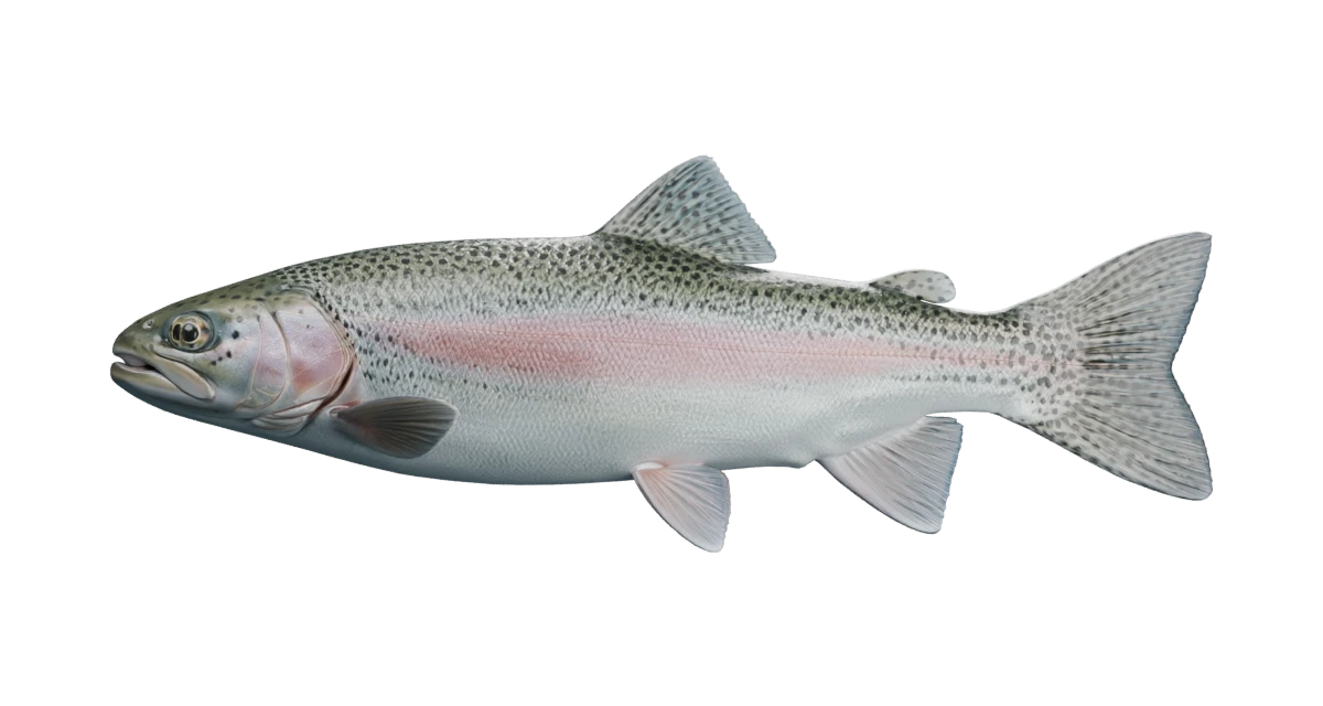
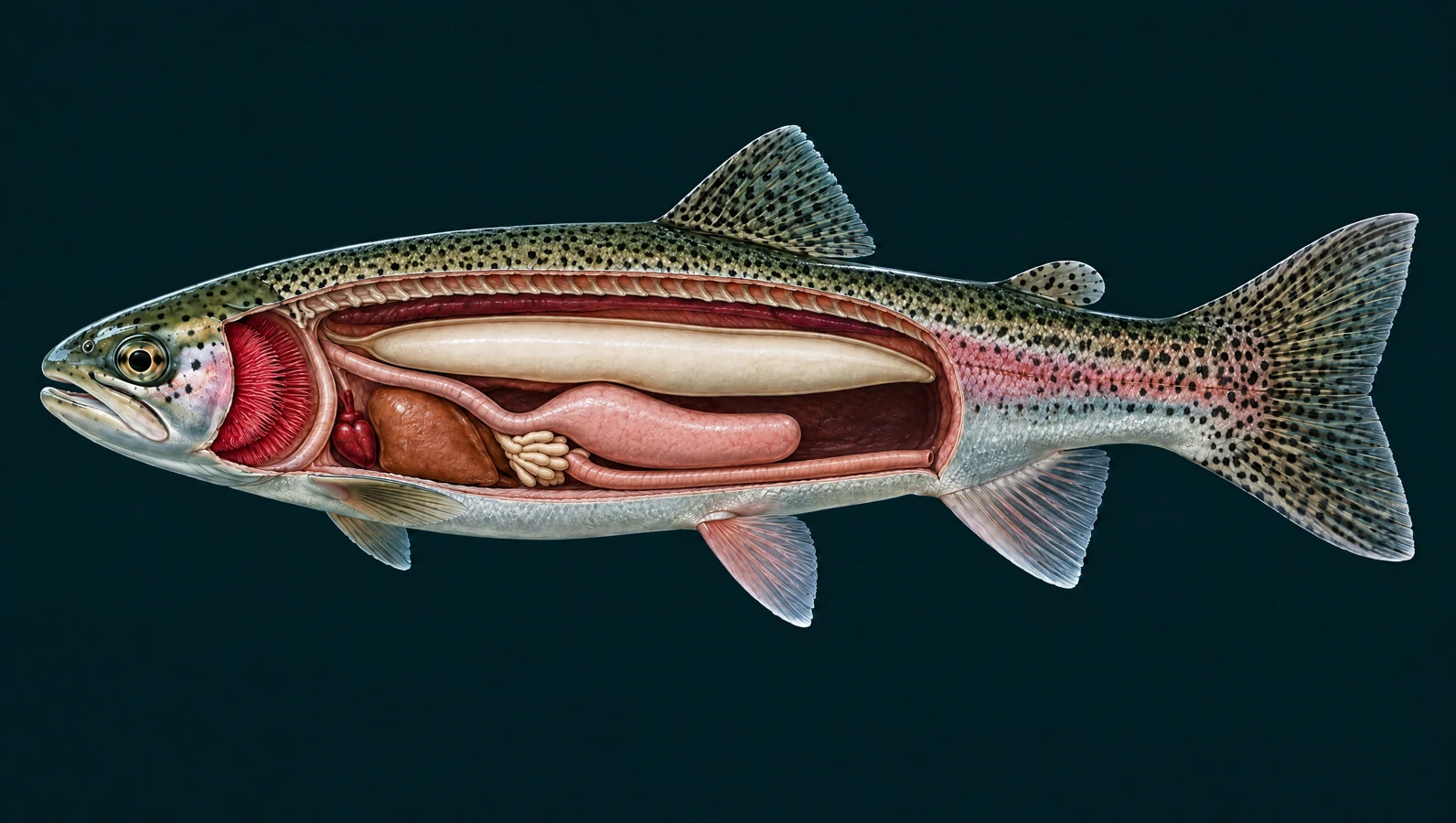
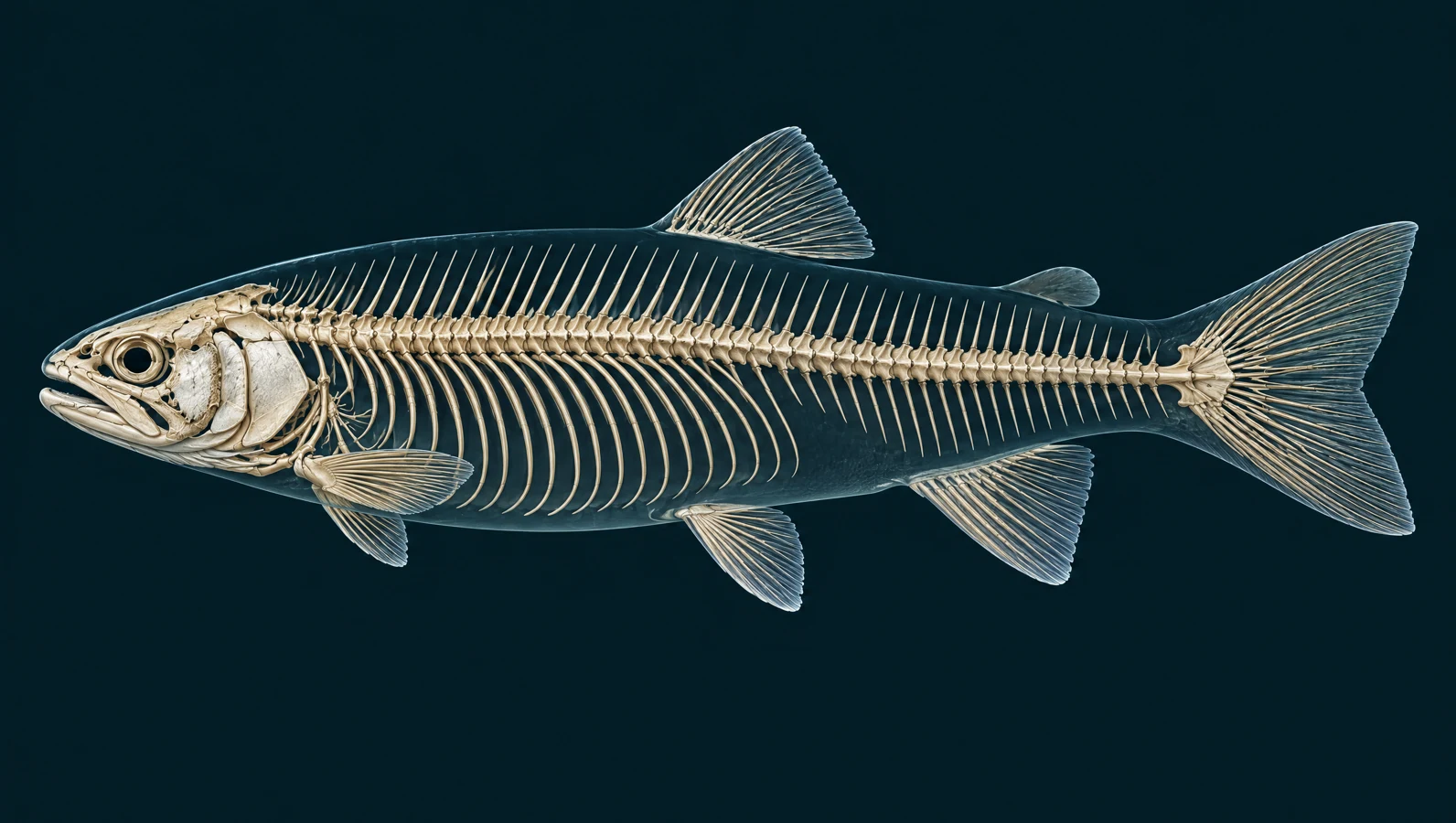
A spotted back, a rosy side, and a body built for life in moving water. Get to know the structures of a rainbow trout, then take a closer look at the fish you meet in Utah.
New: four-step feeding animation. Follow food and water through the fish. Play, pause, or choose a step in the illustrated cutaway.
Fixed-side artist illustrations, not scans. Organ regions and skeletal details are simplified; independent expert review is pending.
The stages separate events for teaching; their timing can overlap in a real fish.
The mouth opens and its cavity expands. Falling pressure draws nearby water and prey inward; a trout may also move toward its food.
Sources: Provini & Van Wassenbergh (2018): suction-feeding hydrodynamics; Jimenez & Camp (2023): vertebral curvature during trout feeding
Water passes through the gill region and leaves behind the gill covers. Gill rakers along the arches help keep food from escaping through the gill passages.
Sources: Provini & Van Wassenbergh (2018): suction-feeding hydrodynamics; Oregon State University / ODFW: Salmonid Dissection Handbook
Retained food moves into the esophagus and stomach. A feeding gulp is not the same as drinking the whole mouthful of water.
Sources: Oregon State University / ODFW: Salmonid Dissection Handbook; Provini & Van Wassenbergh (2018): suction-feeding hydrodynamics
Later, digestive processes break the meal down. The pyloric caeca and intestine help digest and absorb it. This stage takes much longer than the animation suggests.
Aquatic insect larvae and emerging adults are common foods. Insects that fall from the bank can join this moving buffet.
Depending on the water, trout also eat zooplankton, crustaceans, snails and leeches. Available food changes with season and habitat.
Sources: Maine Inland Fisheries & Wildlife: rainbow trout; USGS: Oncorhynchus mykiss species profile
Larger trout may take small fish or fish eggs. They are opportunistic feeders; no single food describes every trout or every lake.
Sources: USGS: Oncorhynchus mykiss species profile; Maine Inland Fisheries & Wildlife: rainbow trout
Back to species guide · Interactive controls become available when JavaScript loads. Read all explanations below.
Takes in food and water. Water entering here passes over the gills before leaving behind the gill cover.
The forward-facing mouth is slightly open in this adult trout reconstruction.
Sources: South Carolina DNR · Fish anatomy; Bureau of Land Management · Junior Explorer fish handbook
Source-checked 2026-09-06. Independent anatomical review pending.
Detects light, shapes and movement. Vision helps fish locate food and notice danger. What a fish can see also depends on light and water clarity.
One of the trout’s two eyes is visible in this side view.
On the water: Seeing a lure is only one part of a feeding response; this diagram does not predict lure success.
Sources: South Carolina DNR · Fish anatomy; Western Australia fisheries · Fish anatomy
Source-checked 2026-09-06. Independent anatomical review pending.
Olfactory openings
Sample dissolved odors. These small openings serve smell. They are separate from the mouth-to-gill breathing route.
The olfactory openings sit just in front of the eye. The marker indicates their general location on the reconstructed surface.
Sources: South Carolina DNR · Fish anatomy
Source-checked 2026-09-06. Independent anatomical review pending.
Operculum
Shields the delicate gills. This bony cover participates in moving water through the gill chamber. Gas exchange happens at the gills underneath.
The curved plate behind the trout’s eye is the cover, not the gills themselves.
Sources: South Carolina DNR · Fish anatomy; Western Australia fisheries · Fish anatomy
Source-checked 2026-09-06. Independent anatomical review pending.
Detects nearby water movement. Small sensory organs along the flank respond to water motion. The highlighted line marks their general course, not a detection radius.
The lateral line and the rainbow’s pink stripe are different features, even where they lie close together.
On the water: Water movement is a sensory cue. It does not establish that a particular vibrating lure works better.
Sources: Western Australia fisheries · Fish anatomy; Utah DWR · Fish identification in the 2023 guidebook
Source-checked 2026-09-06. Independent anatomical review pending.
Helps steady the body. The fin on the back contributes to stability during swimming.
Rainbow trout have a rayed dorsal fin and a separate, smaller adipose fin behind it.
Sources: South Carolina DNR · Fish anatomy; Utah DWR · Fish identification in the 2023 guidebook
Source-checked 2026-09-06. Independent anatomical review pending.
Help turn and slow the fish. These paired fins help with maneuvering and braking.
The near-side fin sits just behind the trout’s gill cover. Its partner is on the other side.
Sources: South Carolina DNR · Fish anatomy
Source-checked 2026-09-06. Independent anatomical review pending.
Assist balance and steering. The paired fins underneath help control the fish’s position in the water.
In trout, the pelvic fins are on the belly, well behind the pectoral fins.
Sources: Western Australia fisheries · Fish anatomy; Utah DWR · Fish identification in the 2023 guidebook
Source-checked 2026-09-06. Independent anatomical review pending.
Helps prevent rolling. This single fin along the lower rear of the body contributes to stability.
Find it behind the trout’s paired pelvic fins and ahead of the tail.
Sources: Western Australia fisheries · Fish anatomy; Utah DWR · Fish identification in the 2023 guidebook
Source-checked 2026-09-06. Independent anatomical review pending.
Caudal fin
Helps propel the fish. Sideways body bends and tail sweeps push against water to produce forward movement.
Small dark spots extend onto the rainbow trout’s tail.
Identification: Compare tail spotting with body pattern and other features; color alone can be misleading.
Sources: South Carolina DNR · Fish anatomy; Utah DWR · Fish identification in the 2023 guidebook; NOAA Fisheries · Fun facts about fascinating fish
Source-checked 2026-09-06. Independent anatomical review pending.
A small fin with a possible sensory role. Research in rainbow trout found nerve-related gene expression consistent with a role in sensing flow. Its full function is still being studied.
This fleshy fin between the dorsal fin and tail lacks the obvious rays of the larger fins.
Identification: An adipose fin is a useful clue, but it is shared by other trout and some other fish.
Sources: Koll et al. (2020) · Gene profiling in the adipose fin of salmonid fishes; South Carolina DNR · Fish anatomy
Source-checked 2026-09-06. Independent anatomical review pending.
Form a protective covering. Overlapping scales and the skin’s mucus layer help protect a fish from injury and infection.
The surface texture illustrates overlapping scales. Scale size and pattern are not a measured identification key.
On the water: Wet your hands before handling a fish to help protect its mucus layer.
Sources: Western Australia fisheries · Fish anatomy
Source-checked 2026-09-06. Independent anatomical review pending.
Gill filaments and lamellae
Take dissolved oxygen from the water. Water passes across thin gill surfaces. Oxygen enters the blood and carbon dioxide moves out; the fish does not split water molecules to obtain oxygen.
The cutaway shows a gill region. Its microscopic exchange surfaces are explained separately, not drawn to scale.
Sources: Huchzermeyer: Introduction to anatomy and physiology of fish
Source-checked 2026-09-18. Independent anatomical review pending.
Pumps blood toward the gills. Blood travels from the heart to the gills to collect oxygen, then to the body before returning to the heart.
Its small, forward position is below and behind the gill region.
Sources: Huchzermeyer: Introduction to anatomy and physiology of fish; California Department of Fish and Wildlife: trout internal anatomy
Source-checked 2026-09-18. Independent anatomical review pending.
Processes nutrients and produces bile. The liver helps handle absorbed nutrients and store energy. Bile supports fat digestion; it is not a passage that food travels through.
The illustrated liver lies near the front of the abdominal cavity.
Sources: Oregon State University / ODFW: Salmonid Dissection Handbook; Huchzermeyer: Introduction to anatomy and physiology of fish
Source-checked 2026-09-18. Independent anatomical review pending.
Oesophagus
Carries swallowed food into the stomach. After capture, food moves from the throat into this short digestive tube. Most water from the feeding strike leaves through the gill openings instead.
Food and respiratory water take different routes after entering the mouth.
Sources: Oregon State University / ODFW: Salmonid Dissection Handbook; Provini & Van Wassenbergh (2018): suction-feeding hydrodynamics
Source-checked 2026-09-18. Independent anatomical review pending.
Holds food while digestion begins. Acid and enzymes start breaking down a meal. The trout stomach has a curved, blind-ended form, with an outlet leading onward to the intestine.
A trout can digest animal prey; digestion is much slower than a feeding strike.
Sources: Oregon State University / ODFW: Salmonid Dissection Handbook; Verdile et al. (2020): the rainbow trout intestine
Source-checked 2026-09-18. Independent anatomical review pending.
Add surface area for digestion and absorption. These finger-like pouches branch near the stomach–intestine junction. They are part of the digestive system, not eggs, parasites or extra stomachs.
The illustration groups many small pouches into one selectable region.
Sources: Verdile et al. (2020): the rainbow trout intestine; Oregon State University / ODFW: Salmonid Dissection Handbook
Source-checked 2026-09-18. Independent anatomical review pending.
Absorbs nutrients from digested food. Digestion continues and nutrients cross the gut lining into the body. Material that is not absorbed eventually leaves through the vent.
Food passes along the intestine; it does not exit through the gill cover.
Sources: Verdile et al. (2020): the rainbow trout intestine; California Department of Fish and Wildlife: trout internal anatomy
Source-checked 2026-09-18. Independent anatomical review pending.
Gas bladder
Helps the fish control buoyancy. This gas-filled organ reduces how much effort the fish needs to hold its depth. It sits above the digestive organs.
In trout, a pneumatic duct connects the bladder with the foregut. It is not the main organ used to breathe in water.
Sources: Oregon State University / ODFW: Salmonid Dissection Handbook; Huchzermeyer: Introduction to anatomy and physiology of fish
Source-checked 2026-09-18. Independent anatomical review pending.
Helps balance water and salts. Freshwater continually enters the body by osmosis. The kidneys help remove excess water in dilute urine while conserving useful substances.
Kidney tissue extends along the dorsal wall, above the swim bladder.
Sources: Huchzermeyer: Introduction to anatomy and physiology of fish; California Department of Fish and Wildlife: trout internal anatomy
Source-checked 2026-09-18. Independent anatomical review pending.
Cranium
Protects the brain and supports the feeding apparatus. A fish head is a complex assembly of bones. Movable parts around the jaws and gill region help expand the mouth cavity during feeding.
Individual skull bones are grouped into one region in this illustration.
Sources: Jimenez & Camp (2023): vertebral curvature during trout feeding; Sugiura (2026): skeletal tissues in rainbow trout
Source-checked 2026-09-18. Independent anatomical review pending.
Open the mouth to capture prey. Jaw movement works with expansion of the mouth cavity and movement of the body. Rapid expansion helps draw water and food inward.
The mouth is part of a coordinated feeding system; the jaws do not work alone.
Sources: Jimenez & Camp (2023): vertebral curvature during trout feeding; Provini & Van Wassenbergh (2018): suction-feeding hydrodynamics
Source-checked 2026-09-18. Independent anatomical review pending.
Spine
Supports the body while allowing it to bend. A chain of vertebrae carries the body axis toward the tail. Muscles act on this flexible framework during swimming and feeding.
Measurements in rainbow trout show vertebral bending during feeding strikes too.
Sources: Jimenez & Camp (2023): vertebral curvature during trout feeding; Sugiura (2026): skeletal tissues in rainbow trout
Source-checked 2026-09-18. Independent anatomical review pending.
Support the walls of the abdominal region. Slender ribs extend from abdominal vertebrae around the body cavity. Farther back, the tail has a different arrangement of supporting bony projections.
This is a simplified regional view, not a complete inventory of every rib and fine bone.
Sources: Sugiura (2026): skeletal tissues in rainbow trout
Source-checked 2026-09-18. Independent anatomical review pending.
Lepidotrichia
Support the spreading surfaces of the fins. Fine bony rays form flexible fans in the dorsal, paired, anal and tail fins. Supports at the tail base transmit movement into the tail fin.
The small adipose fin is soft tissue and has no bony fin-ray fan.
Sources: Western Australia fisheries · Fish anatomy; Sugiura (2026): skeletal tissues in rainbow trout; Koll et al. (2020) · Gene profiling in the adipose fin of salmonid fishes
Source-checked 2026-09-18. Independent anatomical review pending.
Rainbow trout and ocean-going steelhead belong to the same species, Oncorhynchus mykiss. This explorer shows the freshwater form familiar to Utah anglers. Start with the spotted tail, then compare the whole fish.
The tail helps drive the fish forward. The other fins contribute to balance, braking and steering. Play the swimming illustration to follow their movement, or select each fin at your own pace.
Mouth and gill-cover movements move water across the gills. Dissolved oxygen crosses into the blood there. Routine ventilation can happen without eating. During suction feeding, a trout rapidly expands its mouth to draw prey inward.
Water and prey enter together, then separate. Water leaves through the gill openings; retained food travels into the digestive tract. Follow the four numbered stages in the internal view.
The spine provides a flexible body axis. Ribs support the abdominal walls; delicate rays support most fins. The many bones of the head also take part in feeding, allowing the mouth cavity to expand.
Eyes gather visual information, nares sample odors, and the lateral line responds to water movement. These are different ways of sensing the surroundings. The highlighted line is a location, not a map of how far a fish can detect your lure.
The swim bladder lies above the digestive organs, with kidney tissue along the roof of the body cavity. The heart sits forward near the gills. Select each organ to connect its location with its job.
Look for several identification features together. Then connect what you have learned to a Utah species guide, a water guide, and dated stocking records. A species listed at a lake is not evidence of a recent stocking.
A species listed in a water guide is not evidence of recent stocking. Compare the rainbow trout fishing guide, Utah water guides, and dated stocking records.
External structure names, fins, smell and gill-cover function. General fish biology; not a rainbow trout dissection.
Paired fins, skin and scales, lateral line and gill physiology. Text used as reference; artwork is not reproduced.
Rainbow trout exterior and identification. Identification reference only; use current DWR rules when planning a trip.
Scientific name and freshwater rainbow trout / steelhead life histories.
Introductory explanation of water entering the mouth and passing out over gills.
Body, tail and paired fins in fish locomotion.
Rainbow trout evidence supporting a possible flow-sensing function; not proof of a complete behavioral mechanism.
Trout organ locations and relationships; a reference for the simplified cutaway, not a license for the artwork.
Figure 2: salmonid organ arrangement and digestive tract; gill rakers and organ identification. Used for anatomical layout, not all physiological statements in the handbook.
Trout organs, gill countercurrent exchange, circulation and freshwater salt/water balance. Veterinary training material hosted by WOAH.
General mechanics of mouth expansion and water outflow. The study models sunfish, not rainbow trout; it does not validate the timing of this illustration.
Measured body-muscle deformation and vertebral bending during rainbow trout feeding strikes.
Normal control specimens illustrate vertebrae, abdominal ribs, neural/hemal arches and caudal supports. The exhibit does not depict the phosphorus-deficient specimens.
Rainbow trout digestive tract, pyloric caeca and regional digestive/absorptive function.
Diet changes with fish size, habitat and season. Maine fishing regulations and regional size patterns are not applied to Utah.
Rainbow trout feeding ecology and foods including invertebrates, fish eggs and small fish.
Reference fish and river artwork generated with Higgsfield; exterior mesh generated with Meshy 7 through fal. Optimized, annotated and animated in Blender for Utah Stocked Fish, September 2026. Internal and skeletal illustrations generated with Higgsfield GPT Image 2.5 for this exhibit, September 2026. Generated using the connected provider accounts. No third-party Creative Commons license or expert endorsement is asserted.
The 3D exterior and the fixed-side internal illustrations are simplified artistic reconstructions, not scans. Internal views show selected organ regions; overlapping tissues are separated for clarity. Bone counts, small skeletal elements, vessels, gonads and microscopic gill detail are not represented precisely. Flow arrows explain direction, not measured flow or feeding speed. Biological explanations are source-checked; independent expert review of the illustrations and motion is pending.