Insects from water and land
Aquatic larvae and insects arriving from the bank are common prey. A drifting food supply connects a trout’s feeding to the stream and its surroundings.
Sources: South Carolina DNR · Brown trout
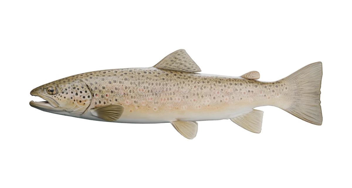
Golden flanks, haloed spots and a long predatory jaw. Explore how a brown trout moves, breathes and turns drifting insects or a larger meal into energy.
New: four-step feeding animation. Follow food and water through the fish. Play, pause, or choose a step in the illustrated cutaway.
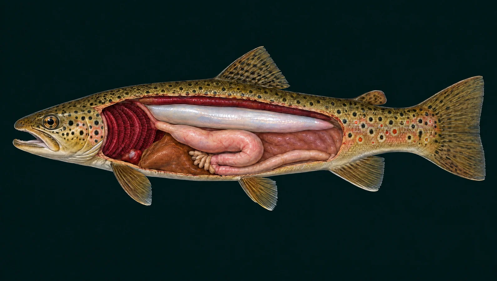
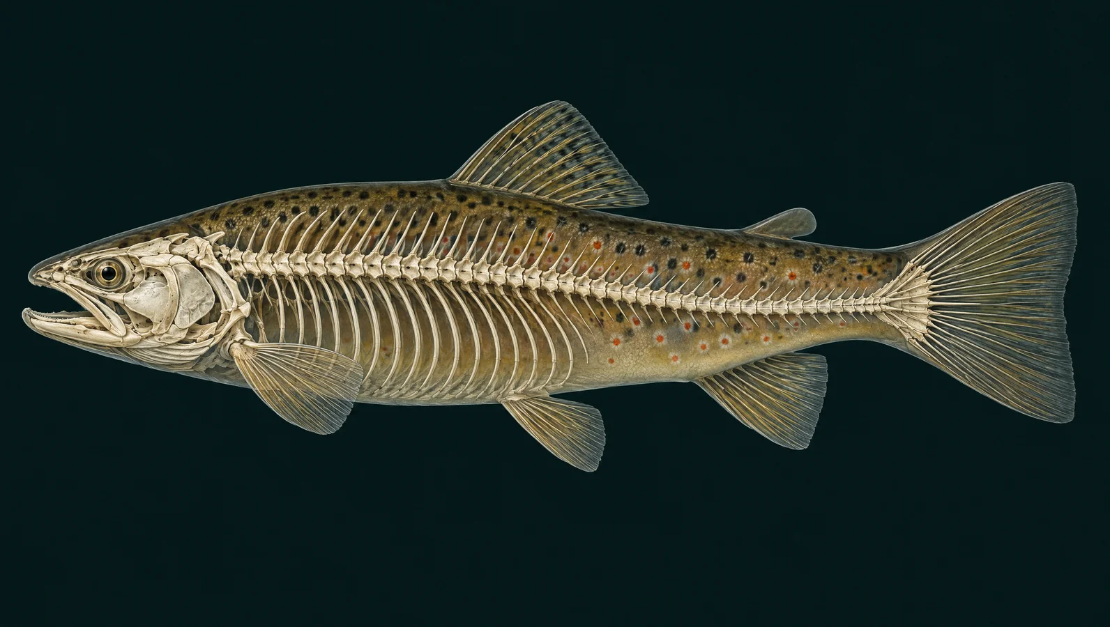
Fixed-side artist illustrations, not scans. Organ regions and skeletal details are simplified; independent expert review is pending.
The stages separate events for teaching; their timing can overlap in a real fish.
The mouth cavity expands, drawing nearby water and food inward. Forward motion can also help close the gap to prey.
Sources: Provini & Van Wassenbergh (2018) · Suction-feeding hydrodynamics
Water continues through the gill chamber and out behind the cover. Gill rakers help retain food; they are omitted from this illustration.
Sources: OSU / ODFW · Salmonid Anatomy & Dissection Handbook; Provini & Van Wassenbergh (2018) · Suction-feeding hydrodynamics
Retained food moves through the esophagus into the stomach. The liver and swim bladder are not part of this food passage.
Sources: Burnstock (1959) · The morphology of the gut of the brown trout
Stomach processing is followed by digestion and absorption along the caeca and intestine. This later stage takes much longer than a feeding strike.
Sources: Burnstock (1959) · The morphology of the gut of the brown trout; Buddington & Diamond (1987): Pyloric ceca of fish, an absorptive organ
Aquatic larvae and insects arriving from the bank are common prey. A drifting food supply connects a trout’s feeding to the stream and its surroundings.
Sources: South Carolina DNR · Brown trout
Browns also take crayfish and fish when available. Diet changes with fish size and the prey present; there is no single standard brown-trout meal.
Sources: South Carolina DNR · Brown trout; Michigan DNR · Brown trout
A northern Utah river study found considerable dietary overlap with Bonneville cutthroat, but brown trout often ate larger items, including fish. That local result is a comparison, not a rule for every water.
Sources: McHugh et al. (2008) · Brown and Bonneville cutthroat trout diets in northern Utah
Back to species guide · Interactive controls become available when JavaScript loads. Read all explanations below.
Takes in prey and water. Expansion of the mouth cavity helps draw nearby water inward. Food can then be retained while water leaves through the gill chamber.
An adult brown trout has a long mouth; the upper jaw commonly reaches beyond the rear of the eye.
Sources: South Carolina DNR · Fish anatomy; Michigan DNR · Brown trout; Provini & Van Wassenbergh (2018) · Suction-feeding hydrodynamics
Source-checked 2026-09-19. Independent anatomical review pending.
Finds movement, contrast and food. Light and water clarity affect the visual information available to a fish.
This eye sits forward of the gill cover on a long, predatory head.
On the water: A visible lure is not automatically a food choice. This model does not predict which color will catch fish.
Sources: South Carolina DNR · Fish anatomy
Source-checked 2026-09-19. Independent anatomical review pending.
Olfactory openings
Sample dissolved odors. These olfactory openings lead to smell tissue. They are not the breathing route to the gills.
The small openings are above the snout, ahead of the eye.
Sources: South Carolina DNR · Fish anatomy
Source-checked 2026-09-19. Independent anatomical review pending.
Operculum
Protects the gill chamber. The bony cover and mouth movements help maintain ventilation. Oxygen exchange occurs on gill surfaces beneath it.
The spotted plate curves around the back of the head.
Sources: South Carolina DNR · Fish anatomy
Source-checked 2026-09-19. Independent anatomical review pending.
Senses local water movement. Sensory cells respond to nearby water motion; the fish does not emit sonar from this line.
The line crosses the golden flank independently of the rows of red and dark spots.
On the water: Vibration supplies information. It does not prove that a particular lure is effective.
Sources: Western Australia fisheries · Fish anatomy
Source-checked 2026-09-19. Independent anatomical review pending.
Contributes to swimming stability. This median fin helps the fish control body orientation.
The larger fin on the back has rays and spots; a separate small adipose fin lies behind it.
Sources: Western Australia fisheries · Fish anatomy; South Carolina DNR · Brown trout
Source-checked 2026-09-19. Independent anatomical review pending.
Help steer and brake. These are paired fins. Changes in their angle help maneuver the fish.
The near-side fin emerges just behind the gill cover; its partner is on the far side.
Sources: Western Australia fisheries · Fish anatomy
Source-checked 2026-09-19. Independent anatomical review pending.
Assist balance and maneuvering. The belly carries a left and right pelvic fin, separate from the single anal fin.
Their abdominal position is a useful contrast with bass, whose pelvic fins sit farther forward.
Sources: Western Australia fisheries · Fish anatomy
Source-checked 2026-09-19. Independent anatomical review pending.
Adds stability near the tail. This single lower fin helps resist unwanted rolling during swimming.
Find it behind the pelvic fins. Its color and pale edging can vary.
Sources: Western Australia fisheries · Fish anatomy
Source-checked 2026-09-19. Independent anatomical review pending.
Caudal fin
Transfers swimming force to water. Alternating body bends and tail sweeps contribute to forward thrust.
A nearly square or slightly concave tail with few or no spots helps distinguish many adult browns from rainbows.
Identification: Combine tail shape with the mouth, body pattern and head markings; a single feature is not enough.
Sources: Western Australia fisheries · Fish anatomy; Michigan DNR · Brown trout
Source-checked 2026-09-19. Independent anatomical review pending.
A small fleshy fin behind the dorsal. It lacks the obvious supporting rays of the larger fins. Its full role in fish biology remains under study.
The small rounded fin may carry dark or reddish pigment. It is not a second rayed dorsal.
Sources: South Carolina DNR · Fish anatomy; South Carolina DNR · Brown trout
Source-checked 2026-09-19. Independent anatomical review pending.
Cover and protect the body. Overlapping scales sit within skin, with a mucus coating that helps protect the surface.
Dark spots and red-orange spots with pale halos decorate the flank. Color varies with environment and life stage.
On the water: Use wet hands and gentle handling to protect the mucus coating.
Sources: Western Australia fisheries · Fish anatomy; South Carolina DNR · Brown trout
Source-checked 2026-09-19. Independent anatomical review pending.
Gill filaments and lamellae
Transfer oxygen into blood. Water passes thin exchange surfaces while blood flows in the opposite direction. The microscopic inset explains countercurrent exchange.
The red arches show a broad gill region; individual lamellae and rakers are not resolved.
Sources: Huchzermeyer · Introduction to anatomy and physiology of fish
Source-checked 2026-09-19. Independent anatomical review pending.
Pumps blood toward the gills. The circulation runs heart → gills → body → heart, with oxygen picked up at the gills.
Look low behind the gills, ahead of the larger abdominal organs.
Sources: Huchzermeyer · Introduction to anatomy and physiology of fish; California Department of Fish and Wildlife · A Trout’s Anatomy
Source-checked 2026-09-19. Independent anatomical review pending.
Processes nutrients and supplies bile. The liver supports metabolism and digestion. Food does not travel through the liver itself.
The rust-brown organ is shown partly lowered so the stomach remains visible.
Sources: OSU / ODFW · Salmonid Anatomy & Dissection Handbook
Source-checked 2026-09-19. Independent anatomical review pending.
Connects the throat to the stomach. Swallowed prey enters this short muscular passage rather than leaving with ventilating water.
Brown-trout anatomical studies describe a short esophagus ahead of the larger stomach.
Sources: Burnstock (1959) · The morphology of the gut of the brown trout
Source-checked 2026-09-19. Independent anatomical review pending.
Holds and begins breaking down a meal. A muscular, curved stomach receives food before it passes onward to the intestine.
Brown trout have a stomach-containing digestive tract. Its J-shaped outline here is simplified.
Sources: Burnstock (1959) · The morphology of the gut of the brown trout
Source-checked 2026-09-19. Independent anatomical review pending.
Add digestive and absorptive area. These blind-ended pouches branch near the stomach–intestine junction; they are not a separate exit for food.
Brown trout have many caeca. The illustration shows a small readable cluster rather than a measured count.
Sources: Burnstock (1959) · The morphology of the gut of the brown trout; Buddington & Diamond (1987): Pyloric ceca of fish, an absorptive organ
Source-checked 2026-09-19. Independent anatomical review pending.
Absorbs nutrients from digested food. After stomach processing, digestion and absorption continue through the gut. Remaining material eventually leaves through the vent.
The posterior path follows the belly. Real gut folds and diameter are simplified.
Sources: Burnstock (1959) · The morphology of the gut of the brown trout; Buddington & Diamond (1987): Pyloric ceca of fish, an absorptive organ
Source-checked 2026-09-19. Independent anatomical review pending.
Helps balance buoyancy. This gas-filled sac reduces the effort required to remain at a particular depth; it is not the stomach.
Brown trout retain a pneumatic-duct connection between this sac and the digestive tract. The fine duct is omitted.
Sources: Burnstock (1959) · The morphology of the gut of the brown trout
Source-checked 2026-09-19. Independent anatomical review pending.
Helps regulate water and salts. Freshwater trout gain water across body surfaces; kidneys help remove excess water while supporting internal balance.
The elongated dark tissue hugs the dorsal body wall above the swim bladder.
Sources: Huchzermeyer · Introduction to anatomy and physiology of fish
Source-checked 2026-09-19. Independent anatomical review pending.
Supports the head and protects the brain. Many connected bones form the fish’s head, including support for its jaws and gill covers.
The broad skull region is shown. Small bones and sutures are intentionally simplified.
Sources: California Department of Fish and Wildlife · A Trout’s Anatomy
Source-checked 2026-09-19. Independent anatomical review pending.
Capture and retain food. Jaw movement changes the mouth opening during feeding. The skull works as an assembly of moving parts.
Fine teeth and joints are illustrative; this is not a dental identification diagram.
Sources: Provini & Van Wassenbergh (2018) · Suction-feeding hydrodynamics; California Department of Fish and Wildlife · A Trout’s Anatomy
Source-checked 2026-09-19. Independent anatomical review pending.
Vertebral column
Provides segmented support for the body. Vertebrae and their connections allow bending while supporting the muscles that move the fish.
The backbone continues into the tail. Its depicted number of segments must not be used to identify species.
Sources: Sugiura (2026) · Skeletal tissues in rainbow trout
Source-checked 2026-09-19. Independent anatomical review pending.
Support the wall around the body cavity. Slender curved ribs occupy the abdominal region; the narrow tail has a different arrangement of bony supports.
This highlighted region stops before the tail. Fine intermuscular bones are not mapped.
Sources: Sugiura (2026) · Skeletal tissues in rainbow trout
Source-checked 2026-09-19. Independent anatomical review pending.
Lepidotrichia
Support the thin fins. Rays spread through the rayed fins and can flex with the fin surface.
The marker selects tail rays. The small adipose fin has no comparable fan of bony rays.
Sources: Sugiura (2026) · Skeletal tissues in rainbow trout; South Carolina DNR · Fish anatomy
Source-checked 2026-09-19. Independent anatomical review pending.
An olive back, warm golden sides and red-orange spots with pale halos introduce Salmo trutta. Look at the whole fish: browns usually lack the rainbow’s broad pink stripe and have much weaker spotting on the tail. The model represents one adult freshwater form; lake fish may look more silver.
The body and tail work together to move water. Paired pectoral fins sit near the head; paired pelvic fins are farther back on the belly. Compare their control surfaces with the stabilizing dorsal and anal fins as you replay the slow swimming illustration.
Opening the mouth and gill covers helps move water across the gills. Dissolved oxygen passes into blood; carbon dioxide moves out. At the exchange surfaces, water and blood move in opposite directions. The little gill inset magnifies that principle rather than depicting a life-size organ.
A brown trout can take insects, crustaceans and fish. During a strike, mouth expansion helps draw prey and water inward. Most ventilating water takes the gill route; retained food follows the esophagus. The four steps separate these routes and slow a rapid event so you can inspect it.
Eyes, olfactory openings and the lateral line collect different kinds of information. A brown trout in cover can use more than one cue to locate a meal or detect danger. Highlighting a sensory structure shows its location, not the distance at which it can detect a lure.
The curved stomach, clustered pyloric caeca and intestine are parts of one digestive system. The liver helps process nutrients, while the long gas-filled swim bladder sits above the gut. These are separate jobs: a fish can regulate buoyancy and digest a meal at the same time.
A fish’s skeleton is not a rigid stick. The vertebral column supports an elongated body, ribs surround the abdomen and rays support the fins. This view maps broad groups, not every tiny bone. See how the ribs end before the tapering tail begins.
Brown trout are introduced fish in Utah. In a northern Utah river study, browns and native Bonneville cutthroat ate many of the same foods, while browns often took larger prey. Explore known waters through the species guide; a species listing is different from evidence of a recent stocking event.
A species listed in a water guide is not evidence of recent stocking. Compare the brown trout fishing guide, Utah water guides, and dated stocking records.
General fin, eye, nostril, gill-cover and lateral-line functions; not a species-specific dissection.
Bony-fish fins, skin, mucus and sensory structures. Marine osmoregulation statements are not applied to freshwater trout.
Figure 2 and the digestive-tract inset establish broad salmonid organ relationships. Used for layout and organ identification, not every physiology statement in the handbook. Artwork not reproduced.
Veterinary training diagrams of trout gills, countercurrent exchange, circulation and freshwater salt/water balance; broad salmonid anatomy, not a measured model of this individual.
Rainbow-trout poster used only to cross-check shared salmonid organ and skeleton relationships. It does not establish this species’ measurements or license the exhibit artwork.
General mouth-expansion and water-outflow mechanics. Sunfish study; no claim that its measured timing describes trout.
Field study comparing opportunistic diets and prey sizes in a northern Utah river. Site-specific results, not a universal diet or proof that every brown trout eats cutthroat.
Normal control specimens show broad salmonid vertebra, rib and fin-support relationships. No transfer of bone counts or phosphorus-deficient anatomy to these illustrations.
Brown-trout identification, diet and use of cover. Regional regulations and size ranges are not applied to Utah.
Adult jaw position, nearly square tail and variable diet. Identification is considered as a combination of traits.
Published abstract supports the short esophagus, muscular stomach, pyloric caeca, intestine and pneumatic-duct connection; no claim of exact counts for the illustration.
Brown-trout neuroendocrine cells and digestive regions; not an uptake-rate experiment, digestion-duration measurement or review of this illustration.
Primary measurements of nutrient uptake in rainbow trout, cod and two bass species support the broad digestive role of caeca. No numerical uptake rate or gut proportion is transferred to brown or cutthroat trout.
Species-specific reference, internal and skeletal artwork generated with Higgsfield GPT Image 2.5. Exterior mesh generated with Meshy 7 through fal, September 2026; prepared for this exhibit in Blender. Generated with the connected provider accounts. No third-party Creative Commons license or expert endorsement is asserted.
An artistic exterior reconstruction and separate fixed-side organ and skeletal illustrations, not anatomical scans. Internal views use broad salmonid relationships; tissues are separated for clarity. Bone counts, vessels, gill microstructure and gonads are not represented precisely. Flow and swimming are explanatory, not measured kinematics. Biological text and broad placement are source-checked; independent expert review of art and motion is pending.